The Image Analysis lab is located in Rooom SN-3097 of the Science
building. There are 2 acquisition set ups in this
room. The first is a macro system consisting of a camera and
light box. The second is a micro system consisting of an
Olympus BX51 microscope. The microscope is caple of both bright field
and epifluorescence imaging.
Each system is equipped with a dedicated PC which is loaded with
Acquisition and Analysis software.
Macro System
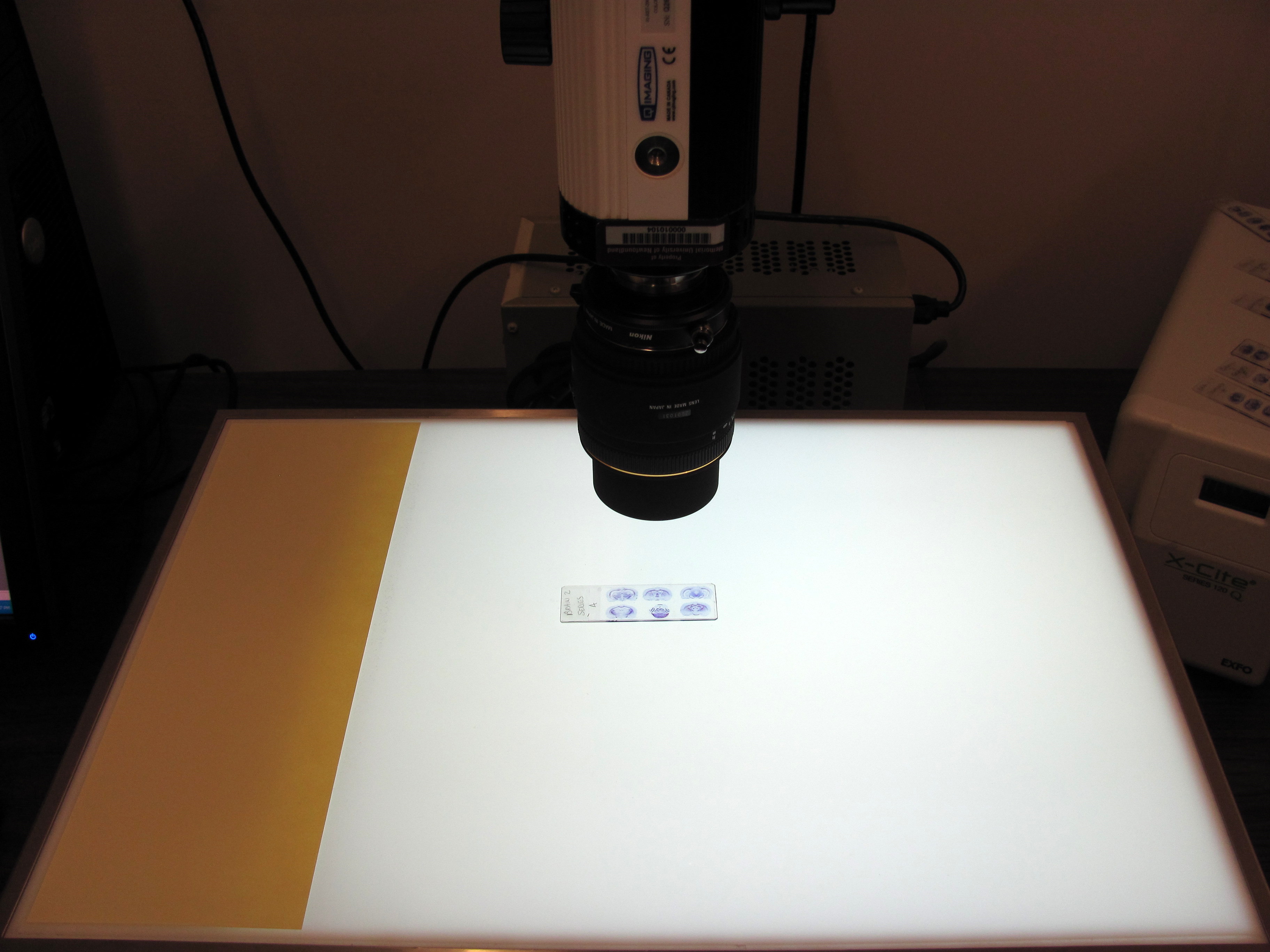
The photo at left shows a microscope slide placed on the
light box under
the camera. The field of view is manipulated by raising and lowering
the camera. You will have to manually refocus the camera
whenever you change the focal length. At maximum
magnification, a coronal rat brain section will fill the
field of view. For higher magnification , the microscope is
needed.
It is a fairly simple system to use. Turn on the camera and
remove the lens cap. Turn on the light box. Turn on the computer and
click in the desktop icon for the acquisition
software,
Qcapture. Click on Acquire/Video Digital to get the control panel
. The
control
panel allows you to"preview" the output from the camera and
"snap" pictures. The quality of the pictures will improve as you become
adept at adjusting focus, aperture, white balance , exposure time...
When focussing, watch the nuber displayed at the bottom of the preview
panel. When this number reaches it maximum value the focus is probably
optimal.
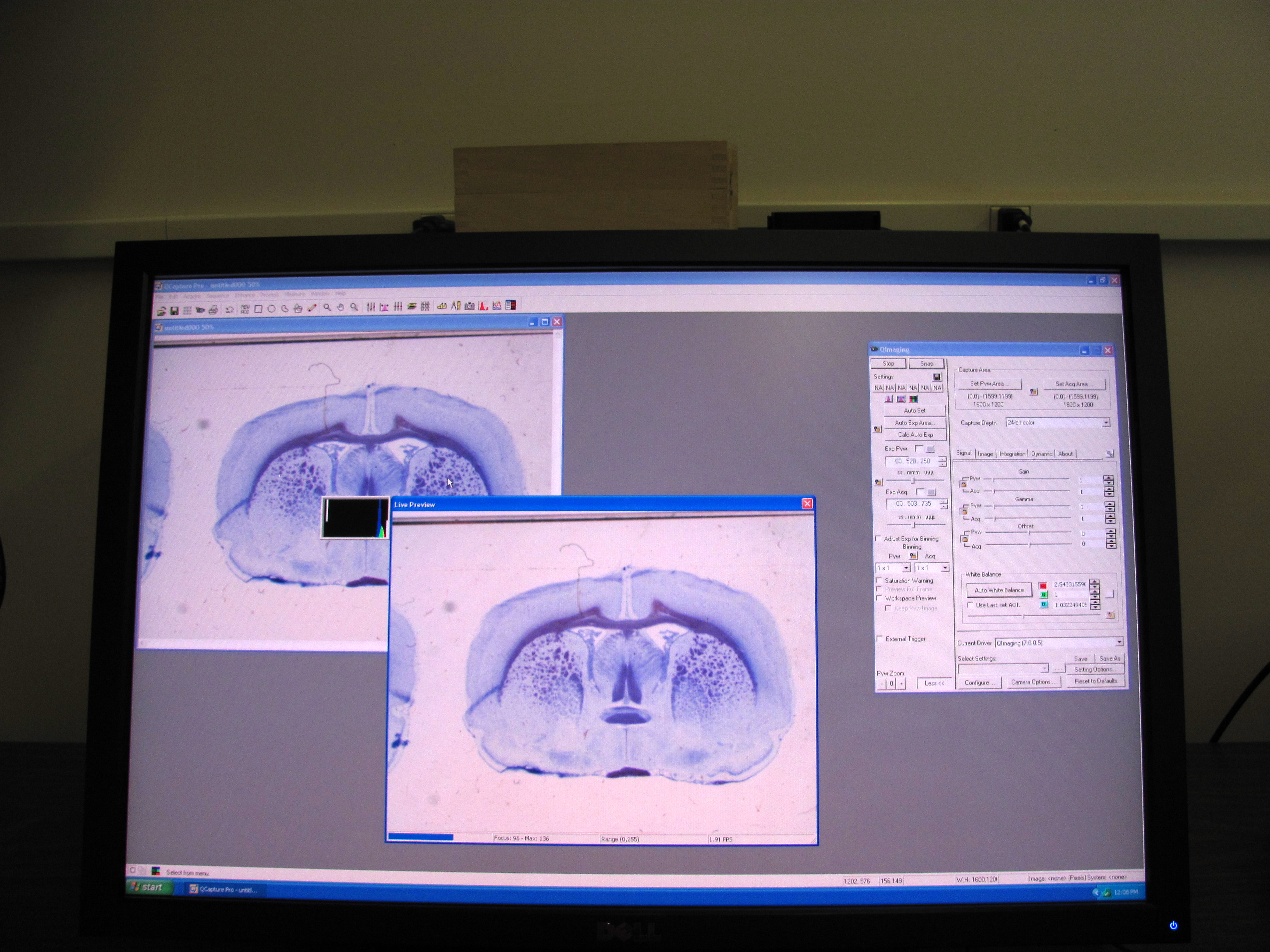
The picture at the right shows the output from the camera and lightbox
above. The camera has been lowered so that a single coronal
section of rat brain almost fills the frame. 3 panels can be seen on
the monitor. the panel at the far right is the control panel . Towards
the center is the preview panel and on the left is the snapshot panel
(the image that is ultimately saved). The control panel allows you to
set the white balance, exposure time...
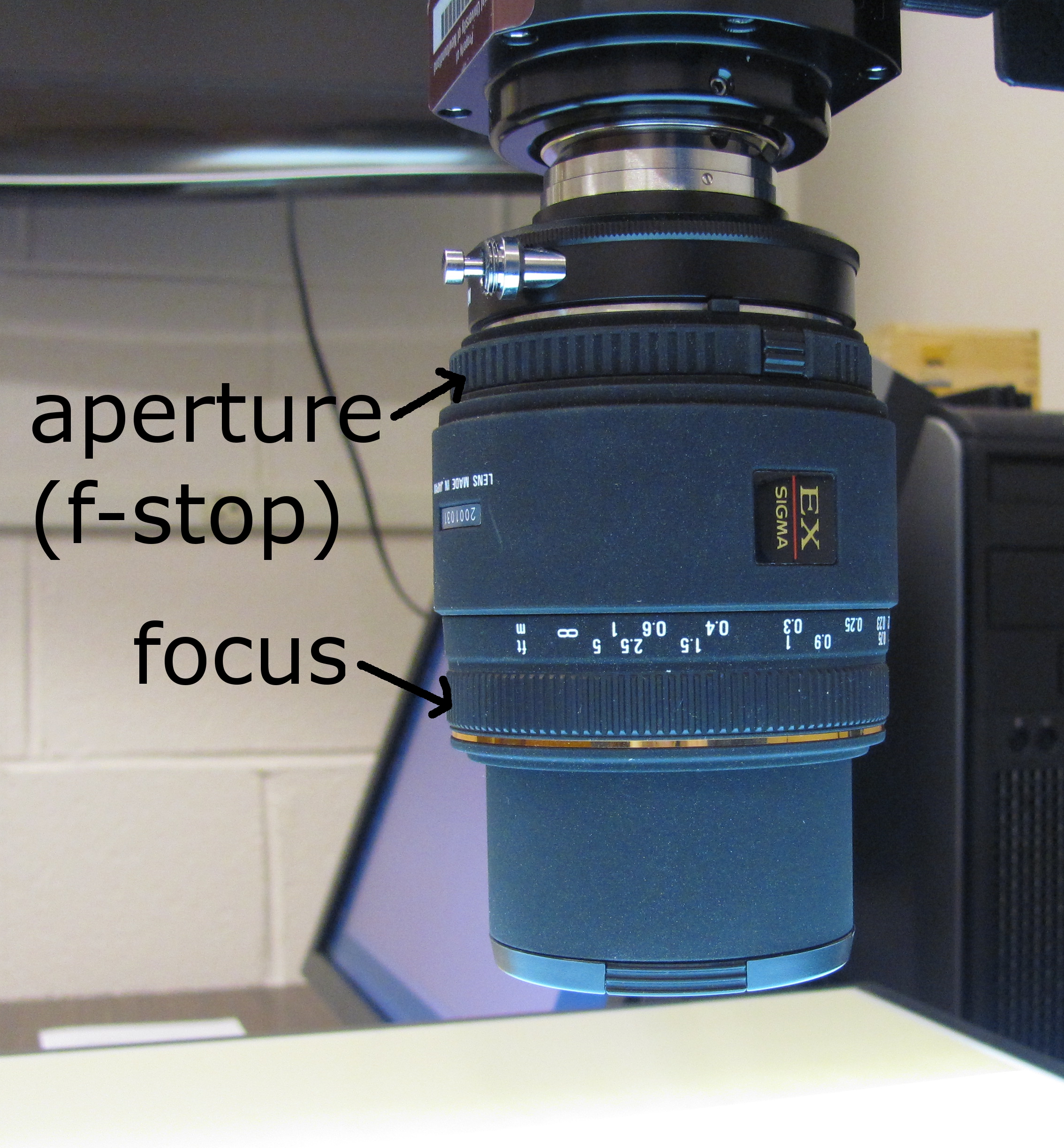
Below is a close-up image of the camera lens showing where to make
adjustments for focus and aperture.
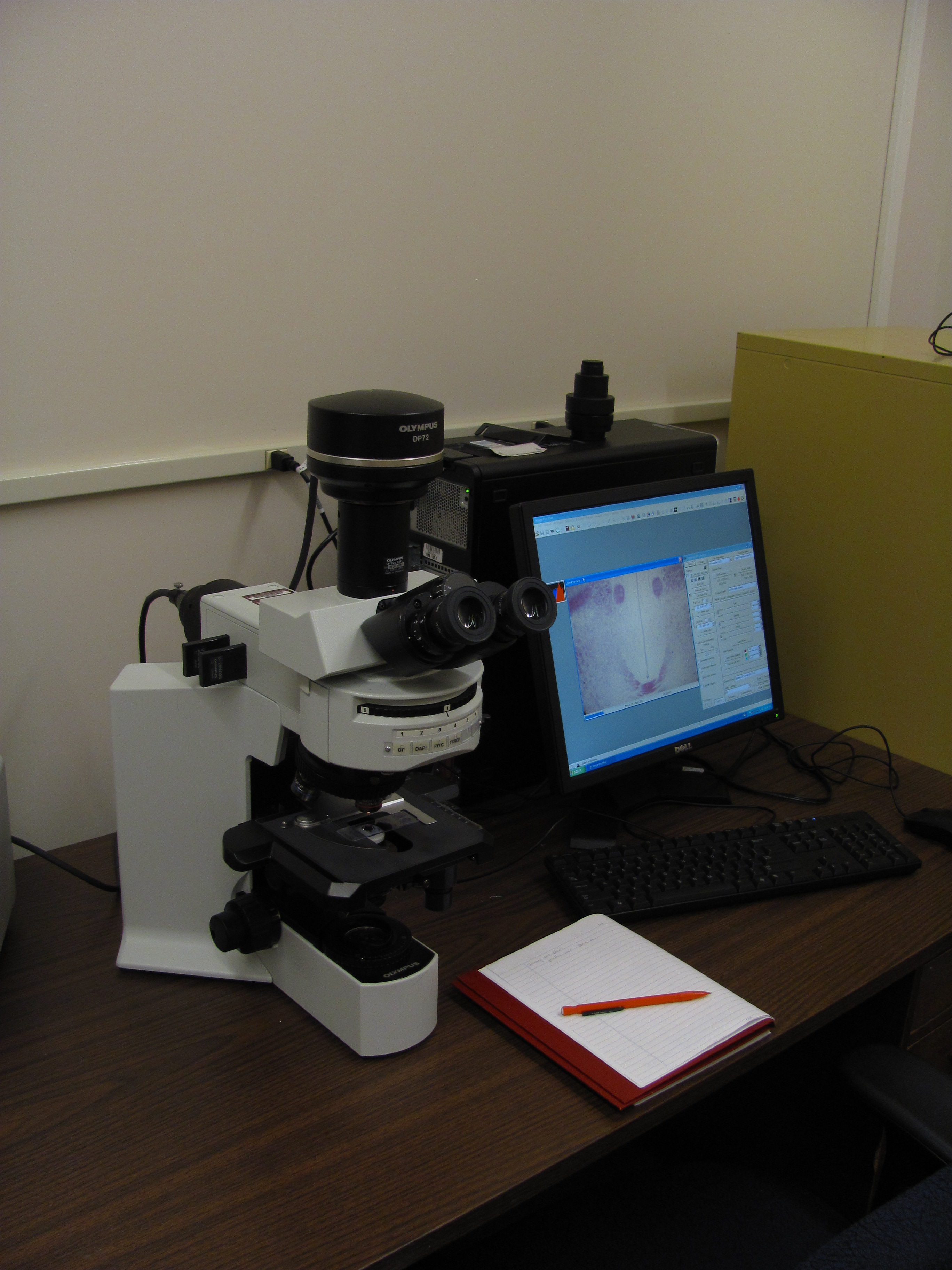
Micro System
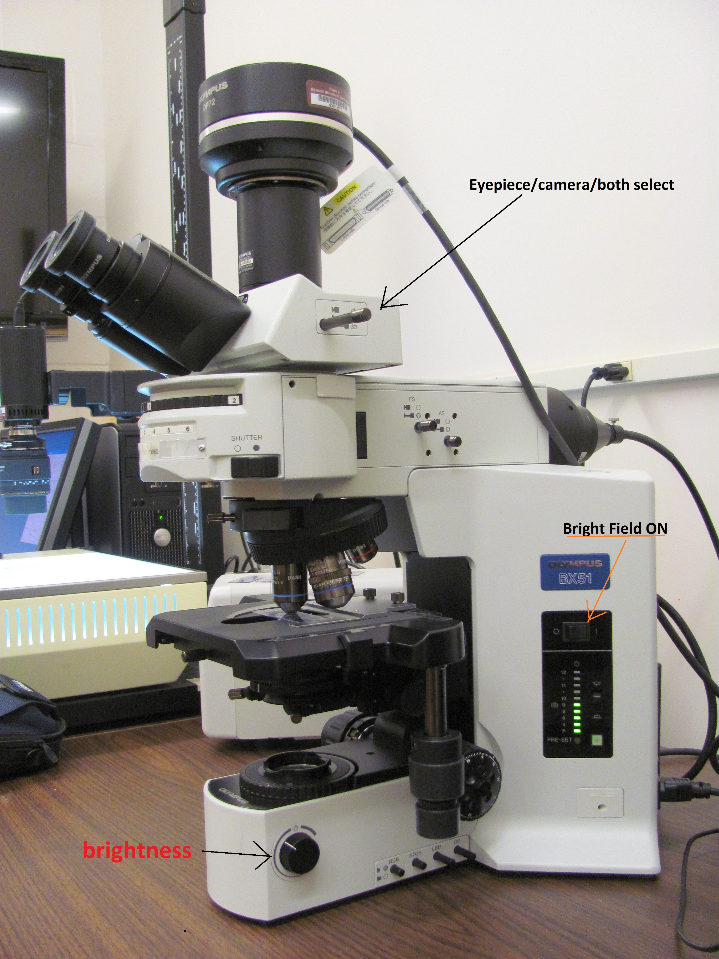 At
left is a photograph of the Olympus BX51 moicroscope with camera.
The slender black rod to the right of the eyepieces
selects whether the image is sent to the eyepieces, the camera or both.
You must select one of the latter two options to view the
image on the computer monitor.
At
left is a photograph of the Olympus BX51 moicroscope with camera.
The slender black rod to the right of the eyepieces
selects whether the image is sent to the eyepieces, the camera or both.
You must select one of the latter two options to view the
image on the computer monitor.
The position of controls for the bright field light source power
and brightness are indicated.
The camera is a very expensive instrument. If you are not
familiar microscopes, seek instruction.
The acquisition software for the Olympus microscope is similar to that
for camera. Double click thet Image Pro Plus icon on the desktop.
Select Acquire/Video Digital Capture. The control panel is almost
identical to the one above. The focus indicator at the bottom of
the preview panel performs like that in the QCapture software.